Function
The facial nerve is, as its anatomy suggests, multifunctional. Its branches control the muscles of facial expression. In addition, the facial nerve is responsible for tear production from the lacrimal gland and taste sensation from the anterior two-thirds of the tongue.
The fine movements of the muscles of facial expression are used continuously to express emotion. In humans the range of facial expressions is extensive and very well developed. Despite being able to verbally express finite and complex feelings and ideas humans still rely on facial expression as a fundamental method of communication.

Motor function
The facial nerve innervates all the branchiomeric muscles of the second branchial arch, in addition to the posterior belly of the digastric, stylohyoid muscle, and the strapedius muscle of the middle ear. Voluntary control over the muscles of facial expression is permitted by two types of motor nerve pathway. Firstly, both cerebral hemispheres supply corticonuclear fibres to the muscles of the upper part of the face. Secondly, the lower muscles of the face are supplied only by corticonuclear fibres from the opposite cerebral hemisphere. However there is a separate involuntary pathway that involves fibres from the reticular formation. Innervation from this pathway inducing smiling or laughing, happens automatically in response to emotion.
Function of the muscles of facial expression
| Muscle of facial expression | Functional movement |
| Occipitalis | Retracts scalp |
| Frontalis | Raises eyebrows |
| Orbicularis Oculi | Closes eyes |
| Corrugator Supercilii | Medially depresses eyebrows and brings them closer together |
| Procerus | Draws skin of forehead down |
| Nasalis | Widens nostrils |
| Orbicularis Oris | Closes and protrudes lips |
| Levator Labii Superioris | Elevates upper lip |
| Levator Anguli | Elevates corners of the mouth when smiling and laughing |
| Zygomaticus | Draws corners of the mouth laterally upwards |
| Risorius | Draws corners of the mouth laterally |
| Depressor Anguli Oris | Depresses corners of the mouth |
| Depressor Labii Inferioris | Depresses lower lip |
| Mentalis | Elevates and protrudes lower lip |
| Buccinator | Compresses cheek |
| Platysma | Depresses mandible and opens mouth |
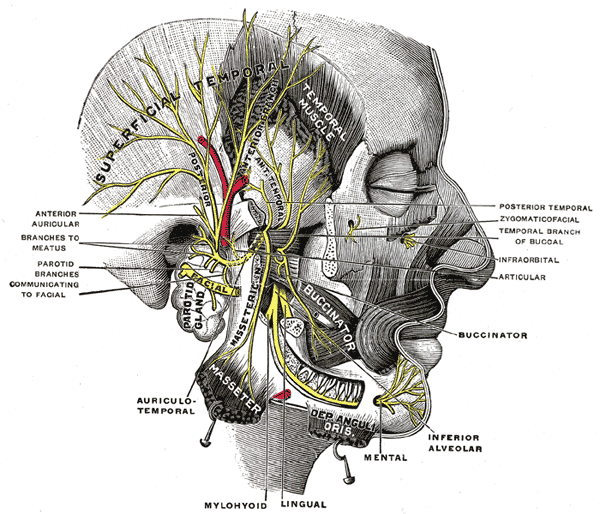
Branches of the Facial nerve and the muscles they supply:
Temporal
Supplies the auriculares anterior and superior, the frontalis, the orbicularis oculi, and corrugator supercilii. The temporal branch acts as the efferent limb of the corneal reflex.
Zygomatic
Supplies the muscles of the zygomatic arch and orbit.
Buccal
Supplies muscles in the cheek and above the mouth.
Mandibular
Supplies the muscles in the region of the mandible but not the muscles of mastication.
Cervical
Supplies the platysma muscle of the neck.
Sensory function
The sense of taste is referred to as a gustatory sense and the tongue is the main taste organ. Sensations of taste from the anterior two-thirds of the tongue travel through the peripheral axons of nerve cells located in the geniculate ganglion on the facial nerve, before passing via the thalamus to the taste area on the cortex. This anterior portion of the tongue is defined as the area in front of the terminal sulcus and primarily has taste buds that detect sweet, sour and salty flavours.
Parasympathetic function
The facial nerve gives off two separate parasympathetic nerves. The first is the greater petrosal nerve and the second is the chorda tympani. The greater petrosal nerve travels through the middle ear and eventually combines with the deep petrosal nerve to form the nerve of the pterygoid canal.
Image copyright details: