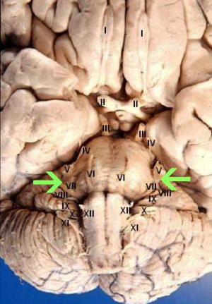
Anatomy
The twelve pairs of cranial nerves include:
- Olfactory
- Optic
- Oculomotor
- Trochlear
- Trigeminal
- Abducent
- Facial (indicated by the green arrows)
- Vestibulocochlear
- Glossopharyngeal
- Vagus
- Accessory
- Hypoglossal
The course of the facial nerve
The facial nerve is made up of an efferent motor root which originates from the facial nerve nucleus in the pons, in addition to a sensory root, arising from the nervus intermedius. The nervus intermedius also contains parasympathetic fibres, these contribute to the autonomic nervous system.
The fibres of the motor root travel posteriorly around the medial side of the abducent nucleus before passing beneath the colliculus facialis in the floor of the fourth ventricle and emerging from deep in the reticular formation of the lower part of the pons. The aspect of the nucleus that supplies the upper facial muscles receives corticonuclear fibers from both hemispheres. However the part of the nucleus that supplies the lower facial muscles only receives corticonuclear fibres from the opposite cerebral hemisphere.
Upon reaching the geniculate ganglion both motor and sensory aspects converge and the facial nerve turns and gives off the greater petrosal nerve, this carries preganglionic parasympathetic fibres. The facial nerve passes through the petrous part of the temporal bone then through the internal acoustic meatus before finally passing through the facial canal. The stapedius muscle division is given off and finally the chorda tympani before exiting the skull through the stylomastoid foramen. The separate branches divide and then go on to innervate specific muscles.
Prior to dividing into its five branches it travels through, but does not innervate, the parotid gland. The chorda tympani carries taste fibres from the anterior two-thirds of the tongue as well as preganglionic parasympathetic fibres towards the submandibular ganglion.
Taste sensation travels from the anterior two-thirds of the tongue, along the peripheral axons of nerves in the geniculate nucleus. These efferent fibres cross the median plane and ascend the ventral posterior medial nucleus of the contralateral thalamus towards hypothalamic nuclei. From here the axons of thalamic cells pass through the internal capsule and corona radiate to the taste area of the cerebral cortex, which is situated in the lower part of the postcentral gyrus.
The parasympathetic nuclei includes both the superior salivatory and lacrimal nuclei. The superior salivatory nucleus receives afferent fibres from the hypothalamus vis descending autonomic pathways. The lacrimal nucleus also receives afferent fibres from the hypothalamus for emotional responses that induce tear production.
Course of facial nerve branches
The temporal branch emerges from the parotid gland and crosses the zygomatic arch and towards the temporal region of the face. The zygomatic branch runs across the zygomatic arch to the lateral angle of the orbit. The largest branch is the buccal branch, which runs horizontally below the eye and around the mouth. The mandibular branch crosses the inferior border of the mandible and passes beneath the platysma muscle of the neck. Finally the cervical branch passes inferiorly of the parotid gland and runs posterior to the mandible.
Location of cell bodies
- Afferent nerve cell bodies are found in the geniculate ganglion for both taste and general afferent sensation.
- Efferent nerve cell bodies are found in the facial motor nucleus in the pons of the brainstem.
- Parasympathetic efferent nerve cell bodies lie posterolateral to the main motor nucleus and are known as the superior salivatory and lacrimal nuclei.
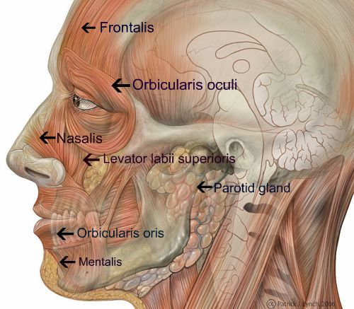
Anatomy of the muscles of facial expression
The image below shows some of the main muscles of facial expression: